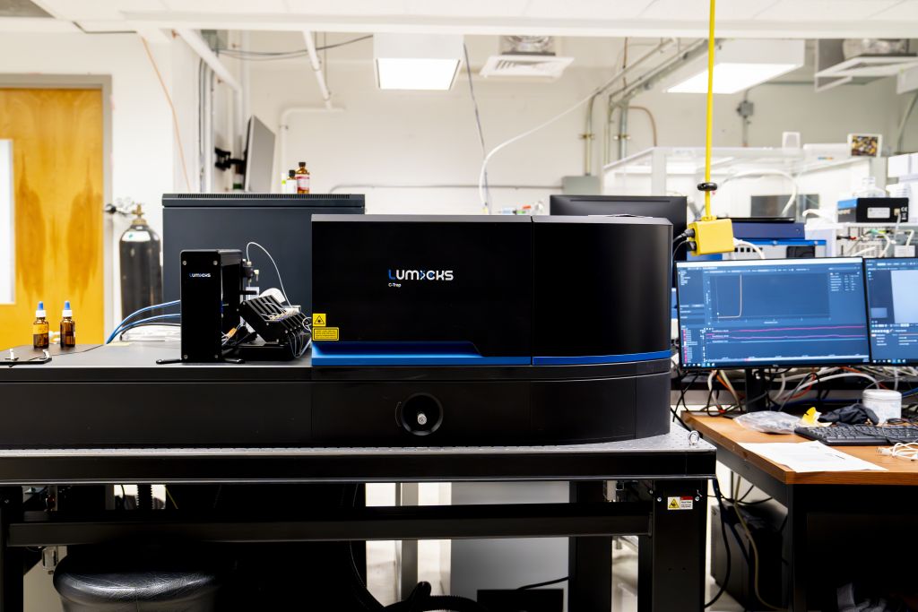
The Lumicks C-Trap2 is a single-molecule force microscope with two optical tweezers correlated with confocal-fluorescence and label-free microscopy
Uses
The multicolor confocal fluorescence imaging allows the visualization of biological processes such as protein/RNA/DNA folding/unfolding kinetics and (un)binding events. It also makes it possible to measure conformational changes of proteins/RNA/DNA by combining the C-Trap with FRET. With its fast 1D scanning capabilities it is suitable for constructs such as DNA/RNA or filaments.
Specifications
- Stable and precise sample manipulation
- A wide variety of visualization capabilities
- High throughput experiment workflow
- Easy and intuitive software package
Details
By shooting a laser through a microscope, a highly focused beam of light is strong enough to trap and hold in place objects such as plastic beads. These beads can be coated to stick to a variety of biomolecules, such as proteins, cytoskeleton filaments, DNA, or RNA. Furthermore, the tiniest forces applied to these molecules can be measured as well, giving the user access to a tool not only capable of manipulating biomolecules but also capable of detecting what’s happening to them. The C-Trap2 provides the world’s first dynamic single-molecule microscope to allow simultaneous manipulation and visualization of single-molecule interactions in real time. It combines high-resolution optical tweezers, fluorescence, label-free microscopy, and an advanced microfluidics system in a truly integrated and correlated solution.
Tae-Hee Lee is the scientific director of the Ctrap2 single molecule biophysics equipment