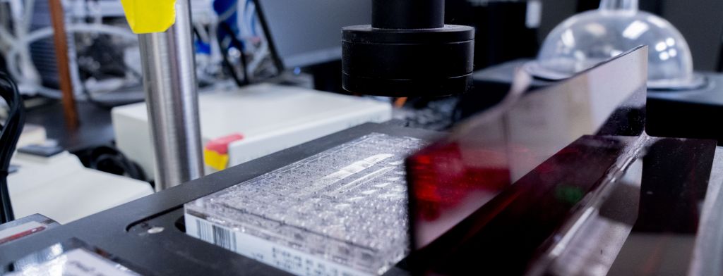
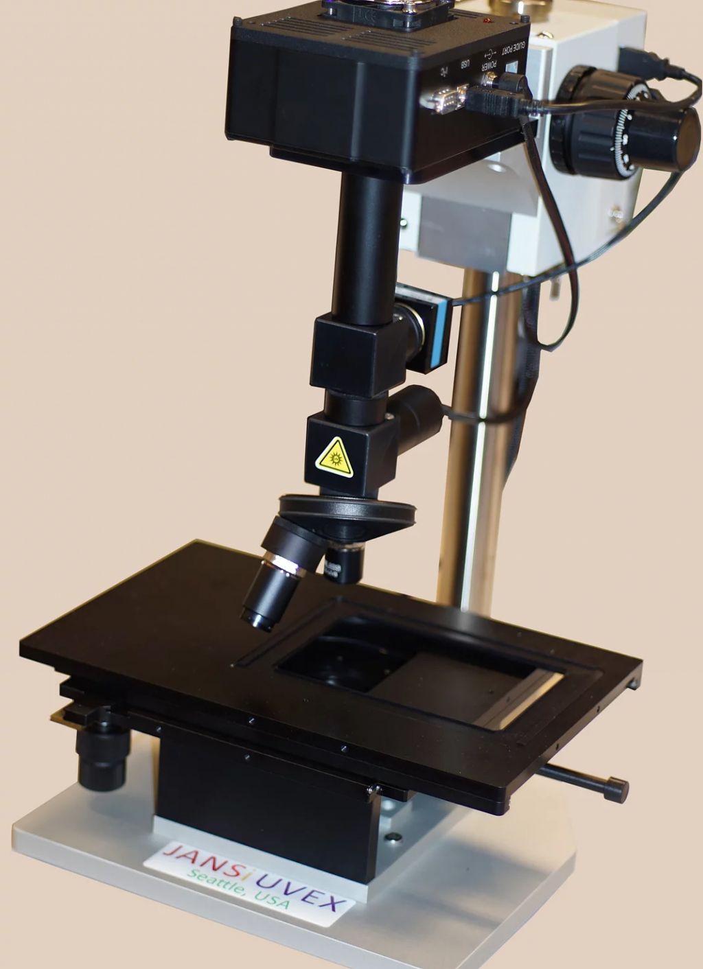
Tryptophan fluorescence at 360 nm, following deep UV excitation at 280 nm, is the basis for this rapid and noninvasive imaging microscope for distinguishing protein and salt crystals in situ
Uses
- Distinguishing protein and salt crystals in situ
Specifications
- Brightfield, UV modes or both modes together.
- Excitation from the top - usable with both sitting and hanging drops
- Camera sensitivity (40% absolute quantum efficiency at 360 nm) to improve detection efficiency
- Highly transmissive (~90% transmittance) filters
- The optical system and camera are highly efficient, giving images with good contrast even with low-intensity deep-UV excitation
- Light sources for excitation and brightfield illumination are compact LED array modules. LED sources are quiet, easier to operate and do not generate any IR radiation that can heat the sample
Acknowledgement
Please acknowledge us as “Penn State X-Ray Crystallography Facility - University Park, PA”