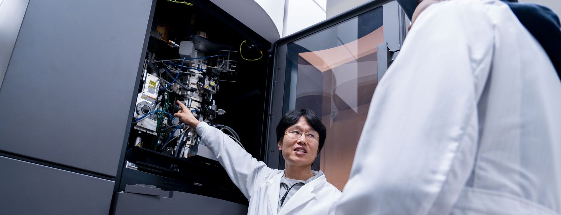
The Huck Cryo-EM Facility at Penn State offers cutting-edge instrumentation and streamlined workflows to support high-impact research. Our facility features two cryogenic Transmission Electron Microscopes (TEMs), a versatile plasma Focused Ion Beam Scanning Electron Microscope (pFIB-SEM), and three semi-automated vitrification systems, enabling fast and efficient sample preparation and imaging. The addition of the pFIB-SEM significantly expands our capabilities, allowing us to support investigations ranging from molecular structures to whole cells and tissues. We proudly serve a diverse user base—from academic researchers to industry partners—and welcome collaborative projects across disciplines.

Cryo-Electron Microscopy Facility
Empowering discovery with automated, high-throughput CryoEM and CryoSEM imaging workflows
News
Stretchy plastics conduct electricity via tiny, whisker-like fibers
A stretchy, conductive type of plastic could help power the next generation of implantable biomedical devices, like longer-lasting pacemakers or glucose monitors, according to Enrique Gomez, professor of chemical engineering at Penn State.
Modern methods in biological research course to be offered in spring 2026
Huck Institutes of the Life Sciences core facilities are offering a new course for spring 2026, Modern Methods in Biological Research, for upper-level undergraduate students and graduate students studying in the life sciences.
Core facilities open house welcomes new researchers
More than 120 researchers attended the first-ever Huck Core Facilities Open House last month, which featured informational posters, opportunities to talk with facilities staff, and even some interactive demonstrations.
September open house to showcase Huck Institutes instrumentation facilities
The Huck Institutes of the Life Sciences will host an open house event on Wednesday, Sept. 24, to advertise the high-tech instrumentation and expert consulting services available in its 11 core facilities.