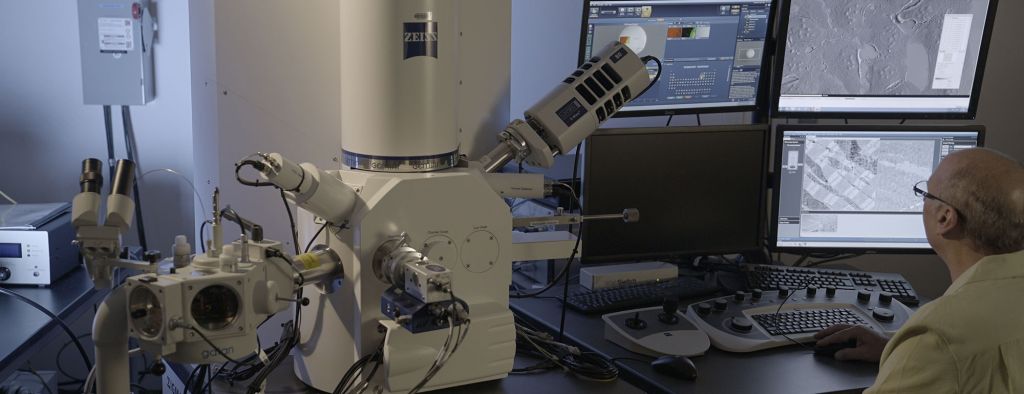
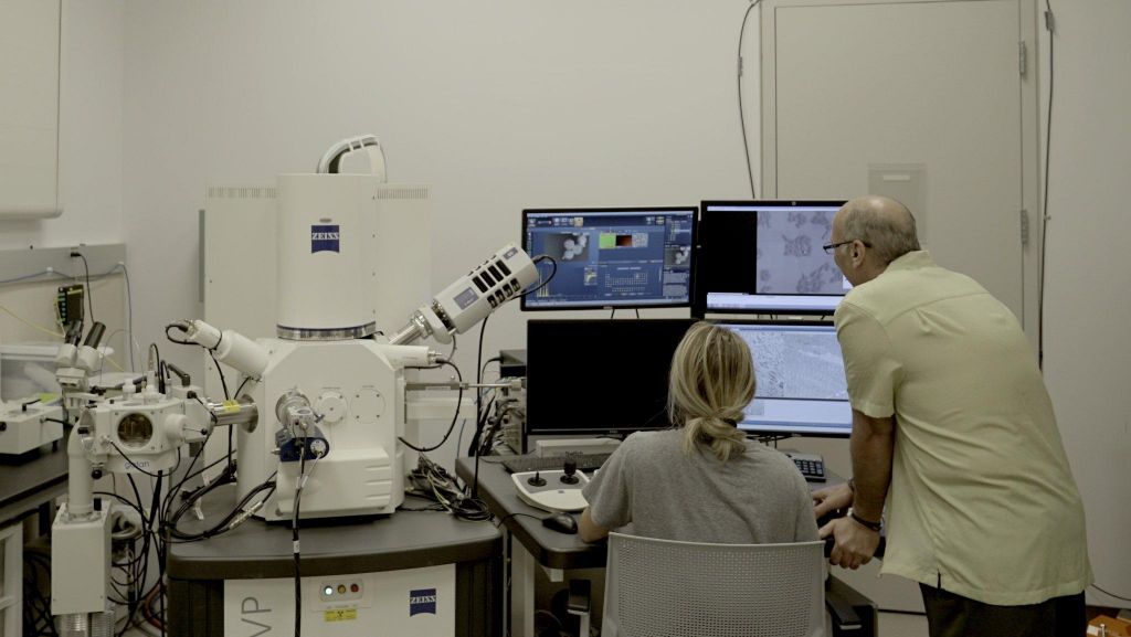
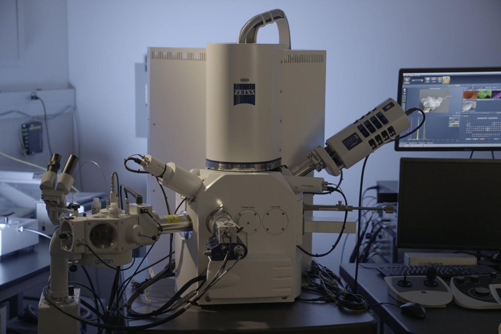
The Zeiss SIGMA VP-FESEM is a variable pressure field emission scanning electron microscope, which allows users to perform high-resolution SEM work for conventional secondary electron imaging, backscattered electron imaging, cryo-SEM, EDS elemental analysis, and 3View reconstruction.
Specifications
- Resolution: 1.3 nm
- Accelerated voltage: 0.1 to 30kV
- Magnification: 12x to 1,000,000x
- 5 Axis Motorized Stage: X=125 mm, Y=125 mm
Details
The scanning electron microscope is an instrument that uses a lens system to focus electrons generated from an electron gun to a fine point at the surface of a conductive sample. The focused beam interacts with the specimen, reflecting electrons which may be absorbed by the sample and subsequently give rise to light (cathode luminescence), electric current, low energy secondary electrons, backscatter electrons, and X-rays. The most useful items to image include the secondary electrons which are generated at the points where the beam interacts with the sample and are subsequently attracted to a detector composed of grid held at a low 50eV positive potential, a scintillator, and photomultiplier tube. The number of secondary electrons are dependent upon atomic identity, topography, and sample orientation at the point of impact. The electron beam is deflected along a straight line on the sample and is terminated at the end of the area set by the magnification selected. It is then returned to the starting point, moved down a short distance, and repeated along a line parallel to the first until an area that is rectangular in shape is scanned. The series of lines constituting the scanned area is referred to as a raster. Deflector coils on the CRT are in synchronization with the scanning coils which affects the SEM beam, but the raster on the CRT is kept constant while the area scanned on the sample can be changed. The ratio of the linear size of the two rasters is the magnification.