Rapid, economical anatomy phenotyping now available to users within and outside of Penn State
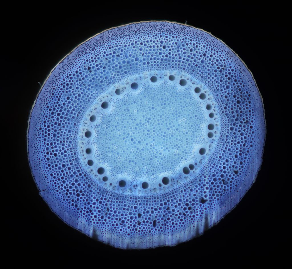
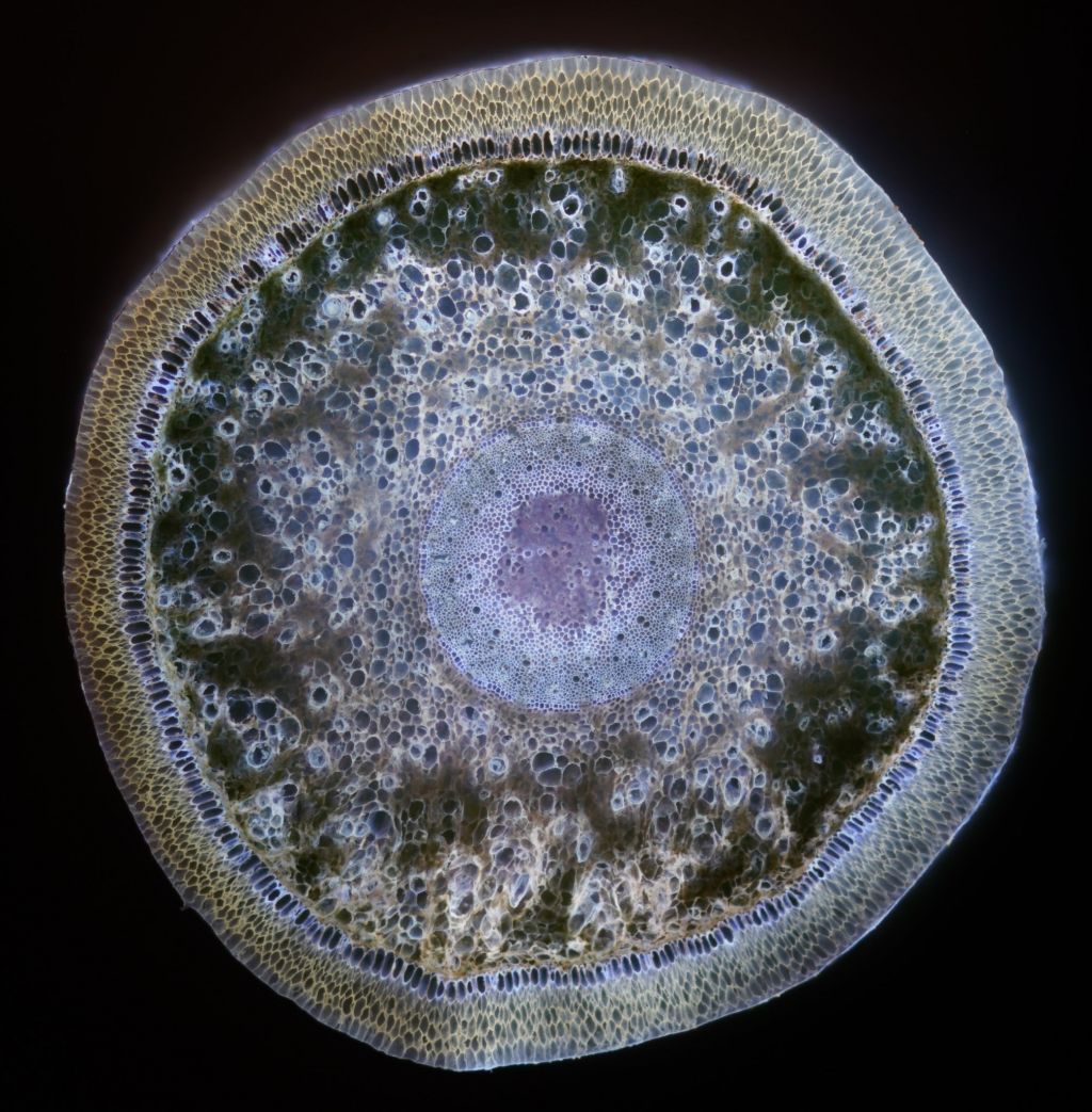
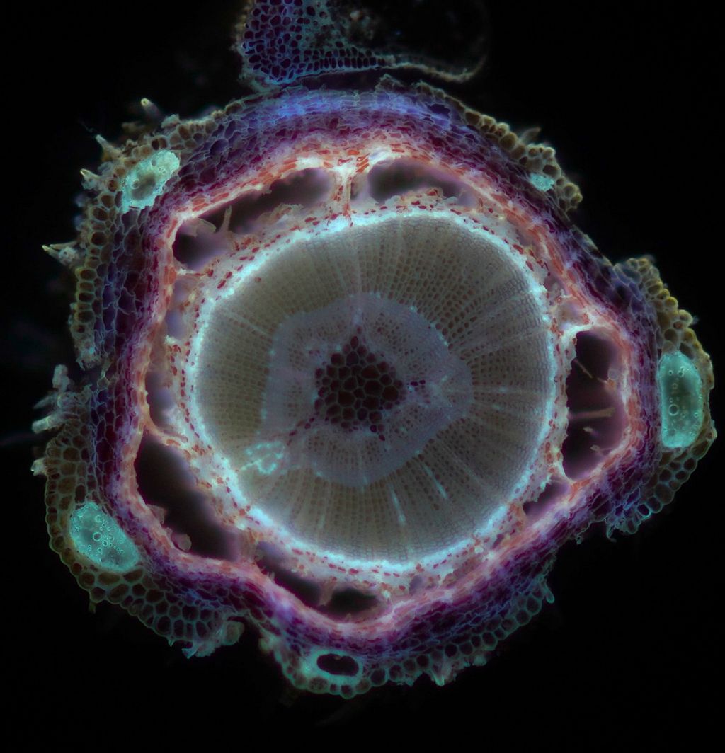
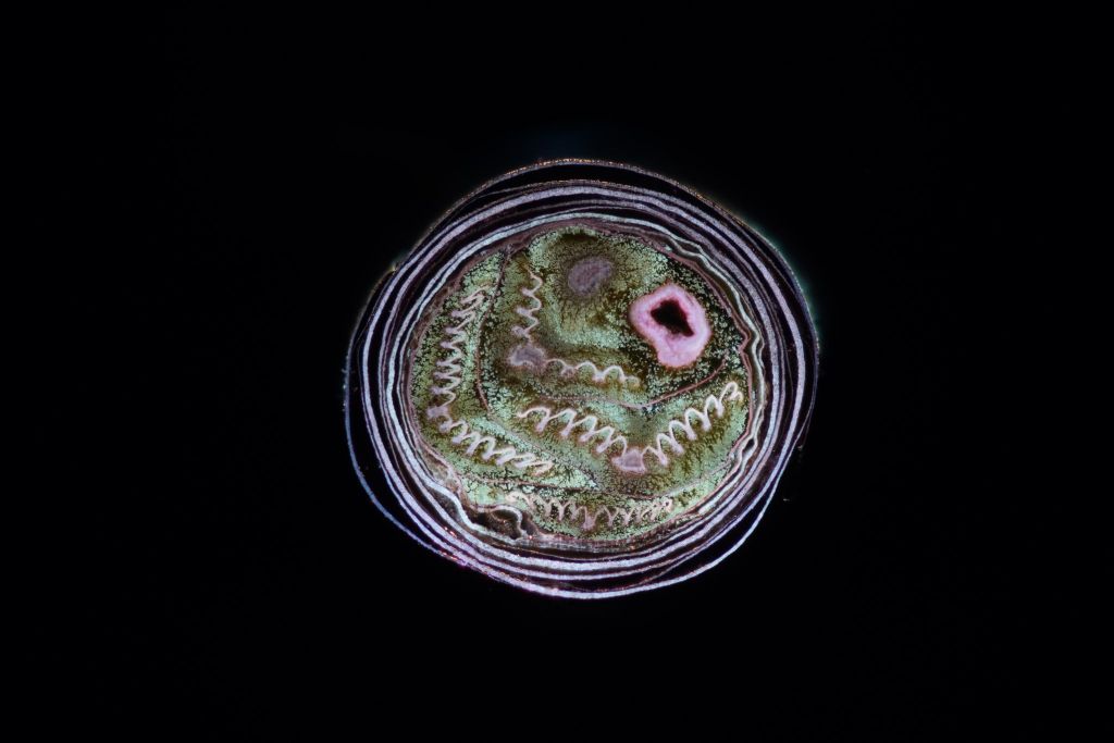
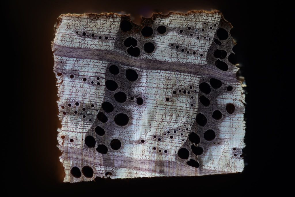
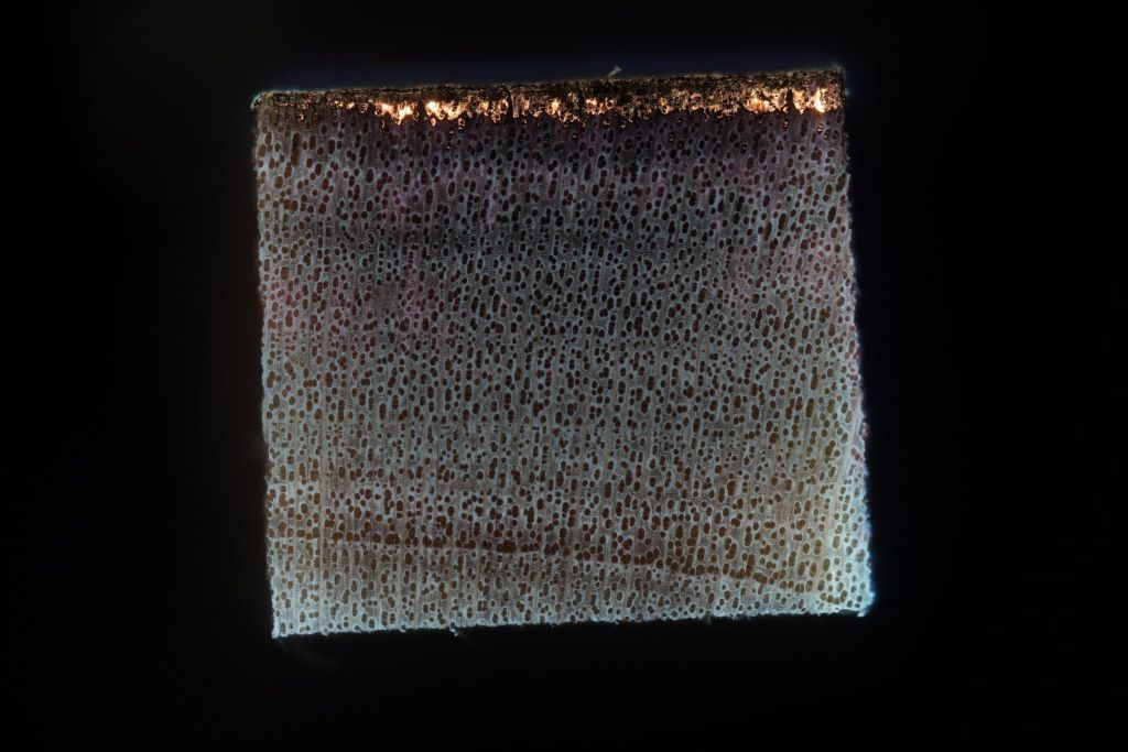
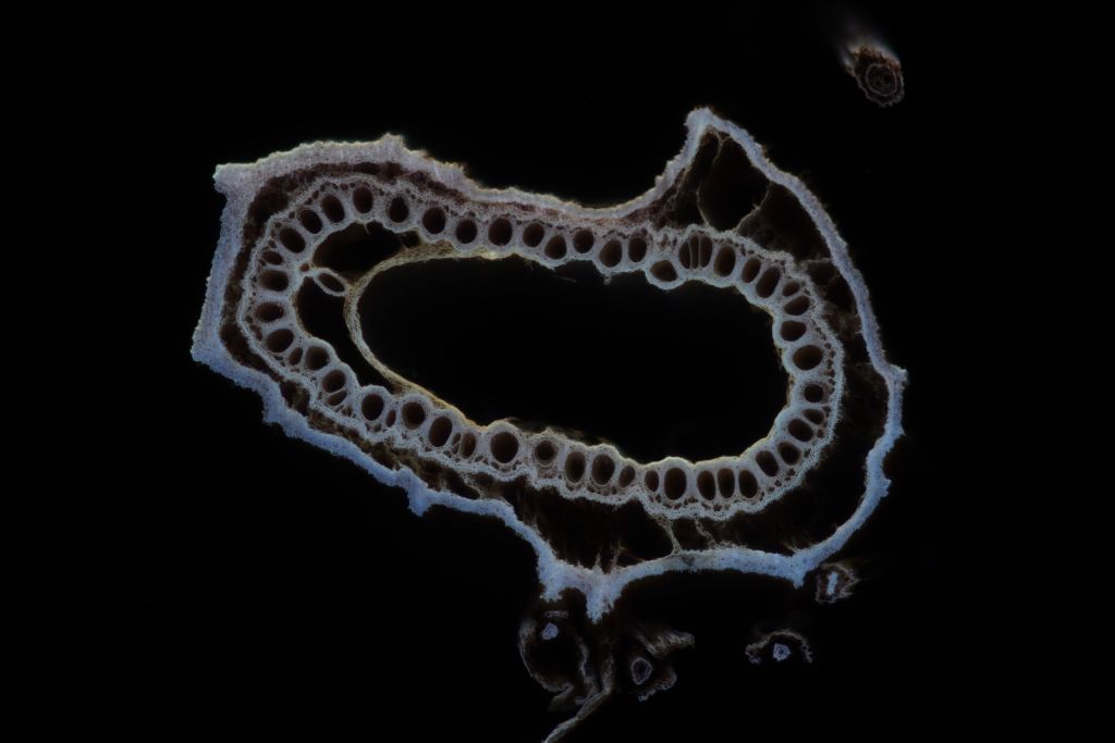
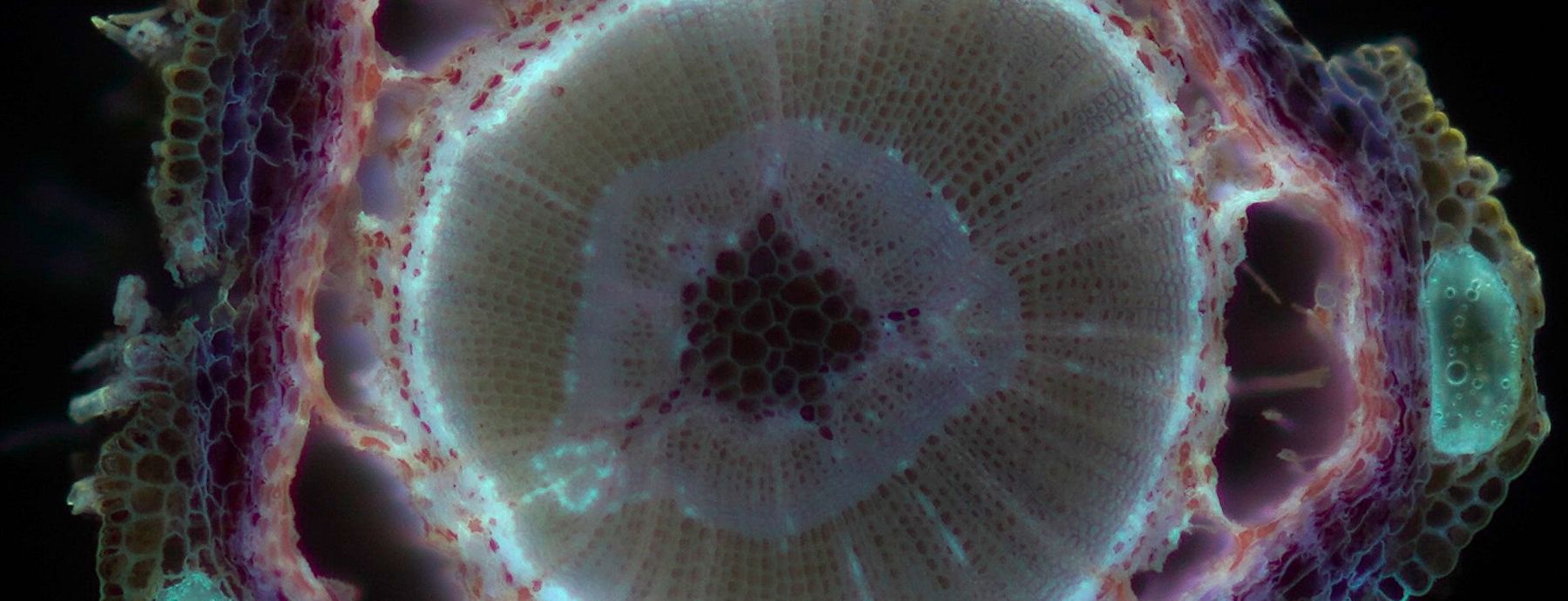
Laser Ablation Tomography (LAT) uses a pulsed laser to section and image experimental samples at tissue/cellular resolution in a single step, significantly reducing time and cost.
No staining, sectioning, or consumables required. Ideal for high-throughput imaging.
Can also generate high-resolution 3D models from z-stacks at a fraction of the cost of microCT.
Why Use LAT?
- Suited for projects with large sample sizes and complex experimental designs where traditional histology is impractical.
- Easy sample preparation. Specimens can be stored long-term in ethanol/water, with no need for fixatives.
- Compatible with a wide range of samples: roots, fungi, insects, and more.
About the Facility
- Two LAT systems available through our core facility.
- Critical point drying (CPD) for sample preparation when necessary.
- Available to researchers affiliated with Penn State as well as those from other institutions.
- Supported by the College of Agriculture, Department of Plant Science, and the Huck Institutes for the Life Sciences.
- Developed at Penn State by Dr. Jonathan Lynch (Dept. of Plant Science)
To Get Started, Contact:
Cody DePew, LAT Facility Manager
cld5311@psu.edu
Our LAT 2.0 system combines high precision hardware to deliver fast, detailed imaging:
- Laser: EdgeWave PX-series 355nm pulsed laser
- 1 mJ energy, 10 picoseconds pulse duration
- Enables athermal ablation (minimal thermal damage)
- Imaging Components:
- Sony Alpha 7R camera (42 MP resolution)
- PRO165LM 3-axis stage
- SCANCube III 10 galvanometer scanner with 103 mm and 160 mm objectives
To get started, we recommend reviewing the following sample prep guidelines:
- Preservation: Store samples in 70% ethanol at room temperature or 4 degrees C.
- Safety: Samples are vaporized. For user safety, they must not be stored in toxic fixatives such as formaldehyde/acetate.
- Sample Types: Most plant tissues, roots, fungal tissues, and small insects. Most free-standing samples can be imaged on LAT.
- Size Guidelines: 5.7mm by 7.7mm field of view, for up to several cm in the z-direction.
Consultation Process:
We begin with a project intake meeting to align on goals, sample needs, and imaging strategy. After submission, typical turnaround is based on scheduling and sample volume.
Data Output:
- Raw image data is provided for your analysis.
- We do not provide full data analysis for most projects, but can recommend tools and consultation, working with you to analyze your data.
The LAT facility operates as a Penn State service center and is open to both internal and external users. Instrument scheduling and additional information can be found on our iLab page. Visit the “PSU Core Facilities” tab at http://psu.corefacilities.org/ and click on “Laser Ablation Tomography” under the College of Agriculture.
Your samples can be imaged in two ways:
- Full-Service Imaging: Facility staff performs imaging on your samples.
- Self-Service Use: Trained users operate the LAT system independently for a lower cost. After receiving access, users can schedule LAT use during regular business hours.
To gain self-service access, users must:
- Provide a valid EHS (Environmental Health and Safety) certificate for the “Laser Fundamentals and Safety” course at https://ehs.psu.edu/training/ (approx. 20 min).
- Complete hands-on training with LAT facility staff (email to schedule 1.5 hr training session).
Rates
- Hourly rates apply and vary depending on service type (full-service vs. self-use) and institution.
- For unique or complex samples, we will work with you to establish appropriate, project-specific pricing.
For rate inquiries or to discuss your project needs, please contact us directly by emailing cld5311@psu.edu.