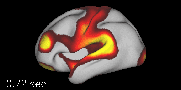
Functional magnetic resonance imaging (fMRI)
fMRI is a non-invasive neuroimaging technique that allows for mapping neural activity across the whole brain. It is based on deoxyhemoglobin concentration changes in brain regions where neural activation occurs. Functional neuroimaging is a predominant method used in studying neural mechanisms underlying mental health disorders.
References:
Tu W, Zhang N (2022): Neural underpinning of a respiration-associated resting-state fMRI network | eLife (elifesciences.org)
Dopfel D, Perez PD, Verbitsky A, Bravo-Rivera H, Ma Y, Quirk GJ, Zhang N (2019): Individual variability in behavior and functional networks predicts vulnerability using an animal model of PTSD | Nature Communications
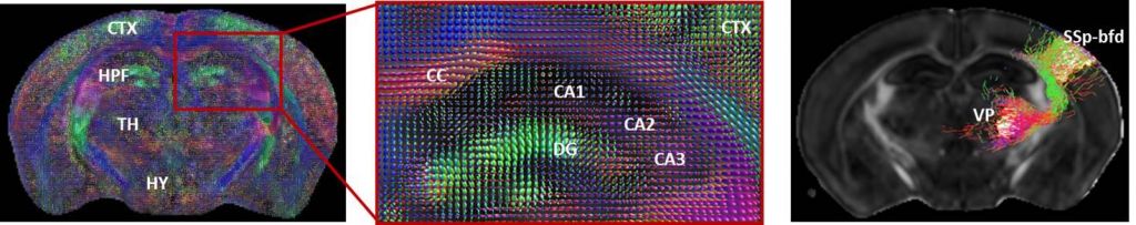
Diffusion magnetic resonance imaging (dMRI)
Diffusion magnetic resonance imaging (dMRI) is one of the most widely used non-invasive techniques that traces diffusion of water molecules along the axons to map the structural networks in the brain. dMRI provides multi-level information about the brain microstructural fingerprints including morphology, and brain network properties. Additionally, with the advent of high-resolution dMRI acquisition and tractography methods, dMRI tractography permits simultaneous examination of multiple white matter connections in the entire brain without tissue sectioning reducing the time and cost of the experiments.
Ultrasound imaging
Ultrasound imaging is a non-invasive imaging modality widely used in clinical practices for diagnostics. Because ultrasound imaging is portable, real-time, inexpensive, and can reach deep tissue, there has been a growing interest in using ultrasound for functional imaging by leveraging ultrafast imaging approaches such as plane wave imaging. In addition, low intensity focused ultrasound has recently emerged as a novel tool for neural modulation and drug delivery across the blood brain barrier. Focused ultrasound could also enable sonogenetics, a new technology that share similar function as optogenetics.
References:
Optogenetics/chemogenetics
Optogenetics and chemogenetics are both neural modulation tools for the activation or inhibition of specific types of cells in the central and peripheral nervous systems. These techniques can be applied independently or combined with other neurotechnology (e.g. fMRI).
References:
Zhifeng Liang, Glenn DR Watson, Kevin D Alloway, Gangchea Lee, Thomas Neuberger, Nanyin Zhang (2015): Mapping the functional network of medial prefrontal cortex by combining optogenetics and fMRI in awake rats - ScienceDirect
Zilu Ma, Qingqing Zhang, Wenyu Tu, Nanyin Zhang (2022): Gaining insight into the neural basis of resting-state fMRI signal - ScienceDirect
Electrophysiology
Patch clamp electrophysiology enables researchers to directly investigate electrical activity in individual neurons and circuits. Researchers can record neuronal membrane currents and membrane potential, as well as other properties related to the wiring and firing of neurons, following various experimental conditions (ex. genetic manipulations; disease and non-diseased state; in vivo and ex vivo manipulations). Patch clamp electrophysiology can be combined with other neuroscience techniques such as optogenetics and RNA sequencing, positioning it as a versatile tool for basic neuroscience research to more translational applications. This depth of circuit and neuronal manipulation can provide greater information to translational relevant research questions by providing a complexity of analysis not available in clinical research.
References:
Dao, Brockway, Suresh Nair, Sicher, & Crowley (2021): SOMATOSTATIN NEURONS CONTROL AN ALCOHOL BINGE DRINKING PRELIMBIC MICROCIRCUIT IN MICE. Neuropsychopharmacology
Suresh Nair, Dao, Lopez Melean, Griffith, Starnes, Moyer, Sicher, Brockway, Meeks, & Crowley (2022): SOMATOSTATIN NEURONS IN THE BED NUCLEUS OF THE STRIA TERMINALIS PLAY A SEX-DEPENDENT ROLE IN BINGE DRINKING. Brain Research Bulletin
Crowley, Bloodgood, Hardaway, Kendra, McCall, Al-Hasani, McCall, Yu, Schools, Krashes, Lowell, Whistler, Bruchas, & Kash (2016): DYNORPHIN CONTROLS THE GAIN OF AN AMYGDALAR ANXIETY CIRCUIT. Cell Reports
Data mining/data modeling
In neuroscience and neuroimaging fields, various brain data collected from single neurons to an entire brain, from mice to humans, from healthy subjects to various patient groups, and from children to aged populations are shared and disseminated every day. This represents unprecedented opportunities but also great challenges for neuroscience communities. The center has been actively engaged in research that develops and applies state-of-art computational and data science tools, including machine learning and artificial intelligence, to various multimodal neural datasets with the purposes of understanding neural mechanisms of basic brain function, elucidating the structure-function-behavior link of the brain, and finding new biomarkers for brain dysfunction. Our ultimate goal is to advance mental health research by modern data science.
References:
Liu X, Leopold DA, and Yang Y. Single-neuron firing cascades underlie global spontaneous brain events | PNAS
Han F, Chen J, Belkin-Rosen A, Gu Y, Luo L, Buxton OM, and Liu X. Reduced coupling between cerebrospinal fluid flow and global brain activity is linked to Alzheimer disease–related pathology | PLOS Biology.
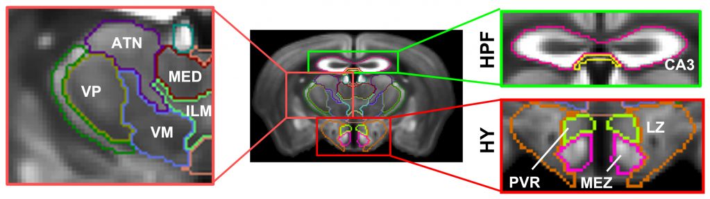
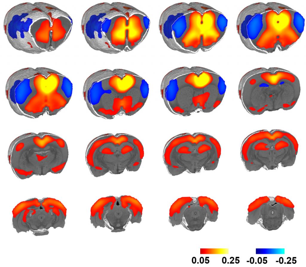
MEMRI
Manganese-Enhancement MRI (MEMRI) is a non-invasive technique that can be used to investigate neurobiological systems with enhanced MR contrast and relatively high spatial resolution using the paramagnetic divalent ion manganese (Mn2+). The chemical similarities between the Mn2+ and calcium (Ca2+) allow Mn2+ to enter excitable cells via voltage-gated calcium channels, the sodium (Na+)/ Ca2+ exchanger, and the Na+/magnesium (Mg2+) antiporter. Therefore, MEMRI technique has the potential to reveal morphological information of the brain as well as to map neuronal tracts by exploring Mn2+ transportation across synapses in the living brain.
In CNMHR, along with the high resolution diffusion MRI and awake functional MRI, we can also determine the activated brain regions due to stimulation, examine axonal transport, brain lesion, and connectivity with high throughput in animal models of human disorders.