Resolution Revolution: Penn State welcomes a new era of atomic-level imaging with cryo EM facility

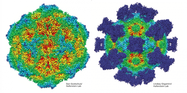
Using extreme cold to arrest fluid samples in motion, cryo EM allows researchers to see proteins, clusters of molecules, and viruses with astounding clarity—to the point where individual atoms may become visible.

Virologist Susan Hafenstein's lab uses cryo-electron microscopy to make high-resolution models of viruses such as the canine parvovirus (CPV) shown here. At left is a CPV capsid, or protein shell, color-coded to indicate surface features. Red areas are depressed and light blue areas are elevated. The capsid at right has been treated with antibodies, shown in dark blue. Binding to the antibodies caused changes in the capsid surface, offering clues to how the virus might be inactivated. IMAGE: DAN GOETSCHUIS (L) AND LINDSEY ORGANTINI (R) / HAFENSTEIN LAB