Jul 02, 2020
Newly acquired Leica SP8 DIVE Multiphoton Microscope enables deep in vivo imaging
Penn State researchers will be able to perform experiments that could not be achieved before. This acquisition will promote research in areas such as neurobiology, cell biology, microbiology, plant physiology, animal sciences and medicine, and beyond.
Amidst the continuing challenges of the COVID-19 pandemic, the Huck Institute’s Microscopy Core Facility has great news to share with the research community. The unit has acquired a Leica SP8 DIVE Multiphoton Microscope with all the “bells and whistles,” thanks to support from the Penn State’s Office of the Vice President for Research and Huck leadership.
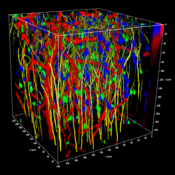
This microscope system enables Penn State researchers to perform deep in vivo imaging of biological and non-biological specimens including live animals, experiments that could not be achieved before. This acquisition will promote research in areas such as neurobiology, cell biology, microbiology, plant physiology, animal sciences and medicine, and beyond.
“I’m excited. This new microscope will benefit Penn State life science community as a whole and bring Penn State’s imaging capability to a new level,” said associate research professor Greg Ning, director of Huck’s Microscopy Facility.
The Leica SP8 DIVE is the first multiphoton microscope with spectrally tunable detection, which combined with its super sensitive hybrid detectors (HyDs) allows for detecting thousands of fluorescent markers. The SP8 DIVE provides maximum penetration depth and best contrast: The new Vario Beam Expander can be tuned for improved penetration depth of more than 1 mm and maximum resolution. This results in the achievement of more contrast and depth for multicolor, deep in vivo imaging.
Some key features of the system are listed below:
• Microscope optics with high transmission efficiency out to 1300nm, optimized for IR range applications.
• Spectra Physics Insight X3 dual beam MP laser: 680 to 1300 nm tunable and 1045 nm fixed.
• Four diode lasers for traditional confocal imaging at 405, 488nm, 552nm, and 638nm
• Conventional FOV scanner and resonant scanner (29FPS at 512x512) for fast biological events.
• Four photon counting HyD’s for external non-descanned detection.
• FALCON – Leica’s fully integrated, and quantitative, FLIM solution
• Prism based spectral detection for traditional confocal imaging, for simultaneous four channel, and up to 8 channel sequential imaging with descanned detectors
• Eight (8) objective lenses
1. HC PL APO 10x/0.40 CS2
2. HC FLUOTAR L16x/0.6 IMM CORR VISIR
3. HCX APO L 20x/0.5 W U-V-I-/D 3.5
4. HC IRAPO L 25x/1.00 W motCORR, WD2.6
5. HC FLUOTAR L 25x/1.00 IMM motCORR VISIR, WD6
6. HCX APO L 40X/0.80 W UVI
7. HC PL IRAPO 40x/1.10 W CORR, WD.65
8. HC PL APO 63x/1.40 OIL CS2
• Application modules
o DIVE Multi-Photon (2P, 3P, SHG, THG)
o FALCON Lifetime imaging (FLIM)
o Full capacity of conventional CLSM function
o LIGHTNING to improve S/N, potentially resolution
o NAVIGATOR for tile scans or multi-position imaging
o FRAP
o FRET
The microscope is located in Room 220 Huck Life Sciences Building.
For more information on the microscope, usage and training, contact Greg Ning at gxn7@psu.edu or visit https://www.leica-microsystems.com/products/confocal-microscopes/p/dive/